Welcome to Midland Cardio Vascular Services Limited
Heartfelt care in the
heart of the Waikato

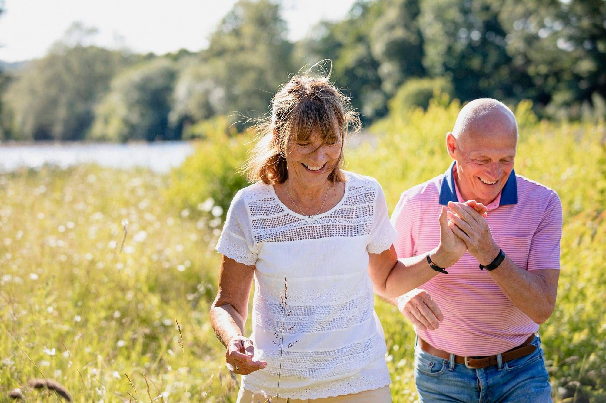
Care that puts you first
At Midland Cardio Vascular Services Limited, we are dedicated to providing top-tier cardiovascular care with a personal touch.
Located within Braemar Hospital in Hamilton, New Zealand, we have proudly served the wider Waikato area and beyond for over 20 years. Our commitment to heartfelt care ensures that each patient receives the utmost attention and quality of service.

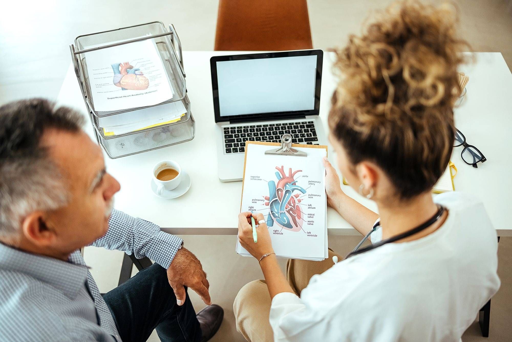
Patient Care
Your well-being is our priority.
We offer compassionate, comprehensive cardiovascular care tailored to your needs. To help you feel confident and prepared for your visit, we provide detailed information on what to expect and support every step of the way.

Visitor info
We welcome support persons and visitors to the Day Stay ward and will ensure their comfort during your stay.
We provide detailed information on visiting hours, amenities, and guidelines to help them feel confident about where to go and how to support you.
Referrers
At Midland Cardio Vascular Services Limited, we value our partnerships with referring healthcare professionals. Your collaboration is essential in delivering seamless, high-quality cardiovascular care.
We provide a streamlined referral process, ensuring patients receive timely, expert care in our state-of-the-art facilities. Our commitment to open communication and comprehensive feedback ensures you are always informed about your patient's progress.
Braemar Hospital
We are proud to operate out of Braemar Hospital, one of New Zealand’s largest and most esteemed private hospitals.
The Hamilton campus, established in 2009, has been continuously expanded and upgraded to maintain its state-of-the-art facilities. Today, Braemar boasts 11 modern operating theatres where around 200 leading credentialled surgeons, physicians, and anaesthetists perform a wide range of elective surgeries, from minor procedures to major operations.
Our Team
At Midland Cardio Vascular Services Limited (MCVS), our dedicated team of highly skilled professionals is at the heart of our success.
Comprising experienced cardiologists, nurses, technicians, and support staff, our team is committed to providing exceptional cardiovascular care. We blend expertise with compassion, ensuring every patient receives personalised and attentive treatment.